Galilei - Dual Scheimpflug Analyzer:
Beyond Topography
The GALILEI™ Dual Scheimpflug Analyzer is a high precision optical system for corneal topography and three dimensional analysis of the anterior eye segment, based on a Revolving Dual Channel Scheimpflug Camera and a Placido Disk.
GALILEI™ combines the advantages of two technologies: Placido imaging furnishes high accuracy curvature data, while Scheimpflug imaging is optimal for precise elevation data.
|
GALILEITM incorporates several important diagnostic modalities. Combining them in a single device not only saves office space and investment cost; it also lays the basis for obtaining consistent, combined diagnostic information from a single set of merged measurement data.
GALILEI integrated functionalities:
- High Resolution Scheimpflug images
- Pachymetry
- Corneal and Lens topography
- 3D Anterior chamber analysis
- Crystalline Lens thickness
- Corneal and lens densitometry
- Pupillometry
|
|
Product Profile
Cataract and refractive surgeons rely on devices that allow for ultra precise imaging to measure corneal architecture and power. The Galilei™ Dual Scheimpflug Analyzer, with its innovative technology, sets a higher standard for diagnosis, biometry, and management of cataract and refractive surgery.
GALILEI™ captures slit images from opposite sides of the illuminated slit, and averages the elevation data obtained from corresponding opposite slit images. This Dual Scheimpflug Imaging technique improves the detection of the posterior corneal surface and provides for outstanding accuracy in pachymetry across the entire cornea, even when the camera is decentered due to eye movements.
Although the resolution of Scheimpflug images is high enough to deliver accurate profile data, it is insufficient to calculate central corneal power (curvature data) with acceptable accuracy. GALILEI™ overcomes this limitation by merging Placido and Scheimpflug data, acquired simultaneously by the two techniques, into a comprehensive single data set. This is essential for obtaining highest accuracy for both elevation and curvature data across the entire cornea.
Near/Far Fixation Target
The GALILEI™ Dual Scheimpflug Analyzer features an adjustable near/far fixation target that allows the examination of the anterior chamber, crystalline lens, and any intraocular lenses at different accommodation states.
The basic technologies inside GALILEI
For an understanding of the functioning principle of the GALILEI Dual Scheimpflug Analyzer and of its inherent advantages over other methods of corneal mapping and anterior segment imaging, it is useful to review the basic concepts of Scheimpflug photography and of Placido Topography. The full advantages of the two complementary methods come to bear when data generated by both methods are combined and merged into a single set of data that describes corneal and anterior chamber geometry.
Dual Scheimpflug Imaging
Scheimpflug imaging differs from conventional techniques in that the object plane, lens plane, and image plane are not parallel to each other, but intersect in a common straight line. The major advantage of the Scheimpflug geometry is that a wide depth of focus is achieved. The Scheimpflug principle has been applied in ophthalmology to obtain optical sections of the entire anterior segment of the eye, from the anterior surface of the cornea to the posterior surface of the lens. This type of imaging allows assessment of anterior and posterior corneal topography, anterior chamber depth, as well as anterior and posterior topography of the lens. |
-
- The angles of the Scheimpflug principle, using the example of a photographic lens.
|
GALILEI captures slit images from opposite sides of the illuminated slit, and averages the elevation data obtained from corresponding opposite slit images. This Dual Scheimpflug Imaging technique improves the detection of the posterior corneal surface and provides for outstanding accuracy in pachymetry across the entire cornea, even when the camera is decentred due to eye movements.
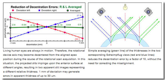
The principal advantage of Dual Scheimpflug imaging is that corresponding corneal thickness data from each view can simply be averaged to compensate for unintentional misalignment, which results in a corrected measurement value at the corresponding location. The dual Scheimpflug imaging principle is independent of inclined surfaces, and thus allows accurate pachymetry without knowledge of the actual decentration of the slit from the apex.
Living human eyes are always in motion even under perfect fixating conditions, and scanning takes time. Therefore, the rotational device axis may become decentered from the aligned apex position during the course of the rotational scan acquisition. In this situation, the projected slits impinge upon the anterior surface of the cornea inclined, resulting in two apparent slit images deviating from each other in thickness. The reciprocal relationship of the dual views allows simple averaging of the corresponding thickness values to correct the values at each of the slit positions. The dual Scheimpflug system has only to take into account decentration and allocate each averaged thickness and posterior height value to its proper location, whereas single Scheimpflug systems additionally have to make estimations on the variable surface inclination for calculating correct thicknesses or posterior heights
Placido Imaging
|
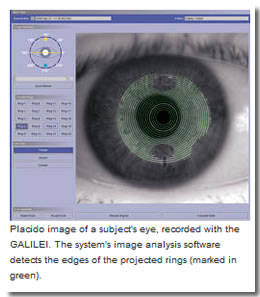
Placido Topography is the classical form of mapping the surface of the cornea with the purpose of determining any deviations of the corneal contour from a perfectly spherical shape. The concentric rings of a Placido Disk are projected onto the cornea to be investigated, and the image reflected back from the cornea is recorded and measured. Geometric analysis of the shape of the reflected rings permits to calculate the steepness, or curvature, of any given point on the corneal surface, and hence to construct a curvature map.
The specific benefit of Placido topography is that it is capable of accurately determining central anterior corneal curvature; a task that is difficult for a Scheimpflug system since elevation differences in the center of the cornea are very small. |
 |
One major shortcoming of Placido topography is that it is measuring reflections on the surface of the cornea only. Therefore the method is not capable of detecting information about the thickness of the cornea.
Merging Scheimpflug and Placido Data
Scheimpflug images furnish precise elevation data of the cornea. Placido images are superior for obtaining accurate curvature data. GALILEI™ combines both.
Although the resolution of Scheimpflug images is high enough to deliver accurate profile data, it is insufficient to calculate central corneal power (curvature data) with acceptable accuracy.
Placido images provide very precise curvature data, even from the central part of the cornea, but they fail to accurately represent small differences in elevation.
GALILEI™ overcomes this limitation by merging Placido and Scheimpflug data, acquired simultaneously by the two techniques, into a comprehensive single data set. This is essential for obtaining highest accuracy for both elevation and curvature data across the entire cornea.
Tech. Features
- Dual Scheimpflug Imaging
Improves the detection of the posterior corneal surface and provides for outstanding accuracy in pachymetry. Details...
- Merging of Placido and Scheimpflug Data
Provides for high accuracy for both elevation & curvature data over the entire corneal surface. Details...
- Near/Far adjustable Fixation Target
Allows examining the anterior chamber, crystalline lens, and any implants under near and far accommodation.
-
Eye Motion Correction
The motion of the eye is tracked based on iris pattern recognition and corrected for to prevent motion artifacts.
Software
|
The GALILEI™ Dual Scheimpflug Analyzer comprises a System Software (operating system and camera controls) and a Basic Applications Software Package. Several additional, mutually independent, optional software packages allow to customize the GALILEI system to the individual needs of the user.
Basic Software Package
Includes corneal and lens topography, best sphere fit elevations, pachymetry, 3D anterior chamber analysis, tools for manual measurements, lens densitometry and opacity map, zonal fit differences, keratoconus screening, and the patient management database
Optional Software Packages
Software Package for separate PC Workstation
Allows data analysis on a separate PC. Measurement data in the patient management database can be accessed directly across a local network.
Accommodation Package
Software package to control the position of the fixation target. The target can be set to different fixed positions, or it can be moved from near to far (or vice versa) during the scan.
Wavefront Package
Includes Anterior Corneal and Total Corneal Wavefront, Modulation Transfer Function, Point Spread Function, and Image Formation on Point Spread Function
|
|