Description
Area Centralis
Volk's Area Centralis is excellent for critically evaluating subretinal neovascularization, atrophic changes of macular degeneration, microaneurysms, diabetic macular edema, proliferative diabetic retinopathy with traction macular detachments, branch retinal occlusion and other retinal conditions.
Area Centralis is properly designed for use within the range of motion of your slit lamp biomicroscope, without compromise of image binocularity or stereopsis.
Primary Application:
High Magnification Viewing and Treatment of the Posterior Pole
Features & Benefits:
• Patented double aspheric glass optics provide enhanced imaging
• Ideal for focal/grid laser treatment
• High magnification image of the posterior pole with expanded field of view
• Available in numerous contact options including our exclusive advanced no fluid (ANF+)
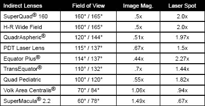
Specifications:
|
lens |
field of view |
image mag |
laser spot |
| Area Centralis |
70°/84° |
1.06x |
.94x |
Available in No Flange Version: VACNF
No Flange (NF) version have a smaller corneal contact area than flanged versions. It is still necessary to use a contact fluid with these versions.
Available in Advanced No Fluid Version: VACANF+
ANF+ flanged version is designed to provide optimal stability without the need for a contact fluid. It may be beneficial to utilize a lubricating fluid to patient comfort.